The eye is a crucial and highly complex sensory organ that enables us to see. It allows us to perceive light, color, and depth. Similar to a camera, the eye processes incoming light to create visual images. Understanding its structure and function also provides insight into how cameras work. Let’s explore the human eye’s structure and functions in detail.
Structure of the Human Eye

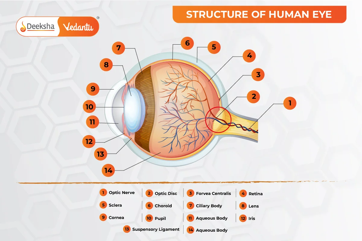
The human eye is roughly 2.3 cm in diameter and resembles a spherical ball filled with fluid. It comprises several parts:
- Sclera: The outer protective white layer of the eye.
- Cornea: The transparent front part of the sclera through which light enters.
- Iris: The colored, muscular ring behind the cornea that controls light exposure by adjusting the size of the pupil.
- Pupil: The opening in the iris that regulates the amount of light entering the eye.
- Lens: Located behind the pupil, it changes shape to focus light on the retina with the help of ciliary muscles, becoming thinner for distant objects and thicker for nearby ones.
- Retina: The light-sensitive layer containing nerve cells that convert images into electrical impulses sent to the brain via optic nerves.
- Optic Nerves: Comprising cones and rods, these nerves transmit visual information to the brain.
- Cones: Sensitive to bright light, aiding in color and detailed vision.
- Rods: Sensitive to dim light, aiding in peripheral vision.
At the junction of the optic nerve and retina, there are no sensory nerve cells, creating a blind spot where no vision occurs.
The eye also includes six muscles: the medial rectus, lateral rectus, superior rectus, inferior rectus, inferior oblique, and superior oblique. These muscles provide various tensions and torques, controlling the eye’s movement.
Function of the Human Eye
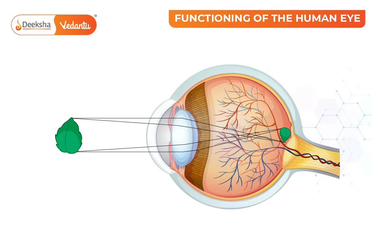
The human eye operates much like a camera, focusing and admitting light to create images. Light rays from distant objects are bent or refracted as they pass through various media—like the cornea, crystalline lens, aqueous humor, lens, and vitreous humor—before landing on the retina.
Refraction, or the change in light direction as it moves through different media, occurs due to the differing refractive indices of these media. This bending of light rays forms an image, which is then received and focused on the retina. The retina contains photoreceptor cells called rods and cones, which detect light intensity and frequency. Millions of these cells process the image and transmit nerve impulses to the brain via the optic nerve.

Here are the refractive indices of various eye components:
- Air: 1.000
- Cornea: 1.376
- Aqueous Humor: 1.336
- Lens: 1.42
- Vitreous Humor: 1.336
Although the image formed on the retina is usually inverted, the brain corrects this inversion. This process mirrors that of a convex lens.
FAQs
The brain processes the electrical signals received from the retina and combines them with information from other sensory modalities to form a coherent visual image. This process involves complex neural pathways and areas of the brain dedicated to visual processing.
The optic nerve carries electrical signals from the retina to the brain, where they are interpreted as visual information. It serves as the primary pathway for transmitting visual information from the eye to the brain.
The blind spot in our vision is caused by the absence of photoreceptor cells (rods and cones) where the optic nerve exits the retina. However, our brains compensate for this blind spot by filling in the missing information based on the surrounding visual information.
Rods and cones are photoreceptor cells located in the retina. Rods are responsible for vision in low light conditions and detecting motion, while cones are responsible for color vision and visual acuity in bright light.
The lens of the eye is flexible and can change shape to focus on objects at different distances. This process, known as accommodation, is controlled by the ciliary muscles surrounding the lens. When we look at objects up close, the ciliary muscles contract, causing the lens to become thicker. Conversely, when we look at distant objects, the ciliary muscles relax, causing the lens to become thinner.
The cornea is the transparent outer layer of the eye that helps to focus light onto the retina. It acts as a protective barrier and also contributes to the eye’s ability to refract light.
The main parts of the human eye include the cornea, iris, pupil, lens, retina, and optic nerve. Each part plays a crucial role in the process of vision. The cornea and lens focus light onto the retina, while the iris and pupil control the amount of light entering the eye. The retina contains photoreceptor cells that convert light into electrical signals, which are then transmitted to the brain via the optic nerve for processing.
Related Topics
- Defects Of Vision And Their Correction
- Pressure
- Electric Potential And Potential Difference
- Full Wave Rectifier
- Zener Diode
- Magnetic Effects Of Electric Current
- Reflection Of Light
- Domestic Electric Circuits
- Bernoullis Principle
- Laws of Motion
- Ohm’s Law
- List of Physics Scientists and Their Inventions
- Spherical Mirrors
- Energy
- Projectile Motion


Get Social