Introduction
The human eye is an intricate and highly sophisticated organ that allows us to perceive the world in all its colors, shapes, and depths. It works by detecting light, focusing it to form images, and sending the signals to the brain for interpretation. Our ability to see is a result of the coordinated function of various parts of the eye, which work together to focus light correctly on the retina.
Structure of the Human Eye
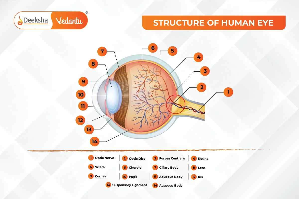
The human eye consists of several parts, each contributing to the overall process of vision. Each part has a specific role in capturing, focusing, and transmitting light.

Cornea
- Description: The cornea is the transparent, convex, outermost layer of the eye. It is the first structure through which light enters the eye.
- Function: The cornea’s primary role is to refract (bend) light rays towards the lens. It provides about 70% of the eye’s focusing power. Since the cornea has a fixed curvature, its refractive power does not change.
- Real-Life Application: When performing LASIK surgery, doctors reshape the cornea to correct vision problems such as myopia, hypermetropia, or astigmatism.
Aqueous Humour
- Description: The aqueous humour is a clear, watery fluid found in the space between the cornea and the lens.
- Function: It helps maintain intraocular pressure, provides nutrients to the avascular parts of the eye (like the cornea and lens), and removes waste.
Iris
- Description: The iris is the colored part of the eye that surrounds the pupil. The color of the iris is determined by genetic factors.
- Function: The iris controls the size of the pupil, which in turn regulates the amount of light entering the eye. In bright light, the iris contracts the pupil to reduce the light entering the eye. In dim light, the pupil dilates to allow more light in.
- Example: If you move from a dark room to a brightly lit area, your iris adjusts the pupil’s size to protect your eye from excessive light, a process called pupillary reflex.
Pupil
- Description: The pupil is the black circular opening in the center of the iris.
- Function: It regulates the amount of light entering the eye by expanding (dilating) in low light conditions and contracting (constricting) in bright light.
- Real-Life Application: Pupil dilation is often performed during eye exams to allow doctors to better view the internal structures of the eye, such as the retina.
Lens
- Description: The lens is a transparent, flexible, biconvex structure located behind the pupil.
- Function: The lens fine-tunes the focus of light that passes through the cornea. It changes shape to focus light onto the retina, a process known as accommodation. The lens becomes thicker to focus on nearby objects and thinner for distant objects.
- Example: The eye’s lens behaves like the lens of a camera, adjusting its curvature to focus light from objects at different distances.
Ciliary Muscles
- Description: The ciliary muscles are tiny muscles located around the lens. They are attached to the lens by suspensory ligaments.
- Function: The ciliary muscles control the shape of the lens, adjusting its curvature for accommodation. When the muscles contract, the lens becomes thicker for focusing on nearby objects; when they relax, the lens becomes thinner for distant vision.
Vitreous Humour
- Description: The vitreous humour is a clear, jelly-like substance that fills the space between the lens and the retina.
- Function: It helps maintain the shape of the eyeball and allows light to pass through to the retina. The vitreous humour also provides support for the retina by holding it in place.
Retina
- Description: The retina is the innermost layer at the back of the eye. It contains millions of light-sensitive cells called rods and cones.
- Function:
- Rods: Responsible for vision in dim light and detecting black, white, and shades of gray.
- Cones: Responsible for detecting color and functioning well in bright light. The retina converts the light that strikes it into electrical signals, which are then transmitted to the brain.
Optic Nerve
- Description: The optic nerve is a bundle of more than a million nerve fibers that connect the retina to the brain.
- Function: It transmits electrical signals from the retina to the visual cortex of the brain, where they are interpreted as visual images.
- Example: The optic nerve acts like a “data cable” that carries the visual information from the eye to the brain.
How the Human Eye Works
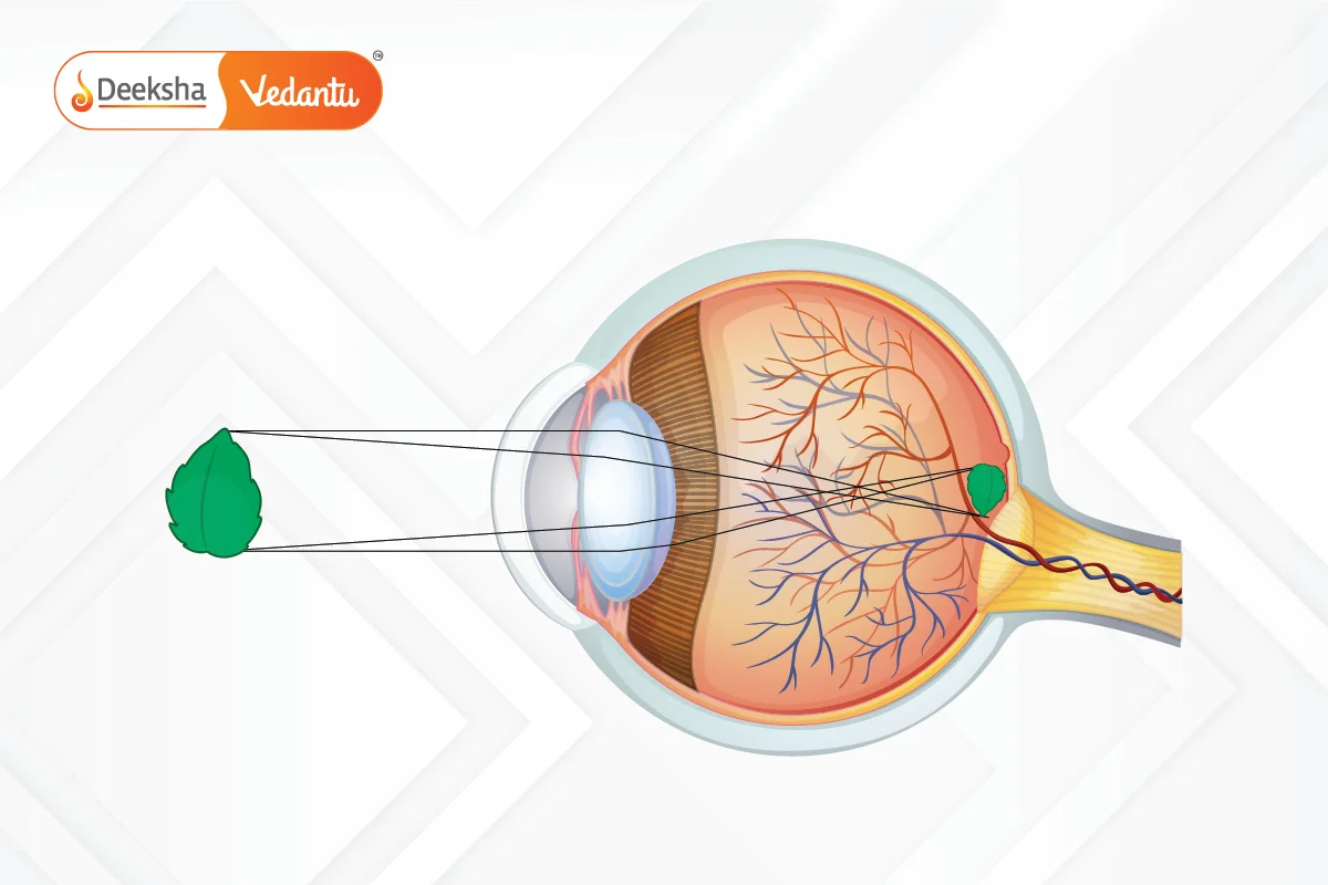
The human eye works by receiving light, focusing it on the retina, and transmitting signals to the brain, which processes them into images. The entire process involves multiple steps:

Step-by-Step Process of Vision:
- Light enters the eye: Light from an object enters the eye through the cornea and is refracted toward the lens.
- Regulation of light: The iris adjusts the size of the pupil to control the amount of light entering the eye.
- Focusing light: The light passes through the lens, which changes shape (due to the action of the ciliary muscles) to focus the light rays on the retina.
- Image formation: The focused light forms a real and inverted image on the retina. The retina’s rods and cones detect the light and convert it into electrical impulses.
- Transmission to the brain: The optic nerve carries the electrical signals to the brain.
- Processing in the brain: The brain interprets the signals and perceives the image as upright and correctly oriented.
Key Concept – Inverted Image:
Although the image formed on the retina is inverted, the brain processes and corrects this image, so we perceive it as upright.
Power of Accommodation
Accommodation is the ability of the eye to change the focal length of the lens to focus on objects at different distances. The ciliary muscles play a crucial role in this process by adjusting the shape of the lens.
How Accommodation Works:
- For distant objects: When you look at distant objects, the ciliary muscles relax, causing the lens to become thinner and flatter, increasing the focal length. This allows light from distant objects to focus properly on the retina.
- For nearby objects: When you look at something close, the ciliary muscles contract, making the lens thicker and more curved, decreasing the focal length. This allows light from nearby objects to focus on the retina.
Limits of Accommodation:
- Near Point: The nearest distance at which an object can be seen clearly without strain is called the near point. For a normal eye, this distance is about 25 cm.
- Far Point: The farthest distance at which objects can be seen clearly is called the far point, which is infinity for a normal eye.
Example of Accommodation:
If you shift your focus from a distant mountain to a nearby book, the ciliary muscles adjust the lens, making it thicker to focus on the text in the book.
Common Defects of Vision
There are several common refractive defects of vision caused by improper focusing of light on the retina. These defects are typically corrected using lenses or other treatments.
Myopia (Near-Sightedness)
- Description: Myopia is a condition where a person can see nearby objects clearly but struggles to see distant objects.
- Cause: The eyeball is too long, or the cornea is too curved, causing the image of distant objects to be focused in front of the retina.
- Correction: Myopia is corrected using concave lenses, which diverge the light rays before they enter the eye, allowing them to focus on the retina.
Example:
A person with myopia may have difficulty reading road signs from a distance. They would need glasses with concave lenses to see clearly.
Hypermetropia (Far-Sightedness)
- Description: Hypermetropia is a condition where a person can see distant objects clearly but struggles to focus on nearby objects.
- Cause: The eyeball is too short, or the lens is too flat, causing the image of nearby objects to be focused behind the retina.
- Correction: Hypermetropia is corrected using convex lenses, which converge the light rays before they enter the eye, allowing them to focus on the retina.
Example:
A person with hypermetropia may need to hold reading materials farther away from their eyes to see clearly. Convex lenses in their glasses will help them focus on nearby text.
Presbyopia
- Description: Presbyopia is an age-related condition in which the lens of the eye loses its flexibility, making it difficult to focus on nearby objects.
- Cause: As people age, the ciliary muscles weaken, and the lens becomes less elastic, reducing its ability to adjust its shape for near vision.
- Correction: Presbyopia is corrected using bifocal lenses or progressive lenses, which help with both near and far vision. Bifocals have two parts: one for distance vision and one for near vision.
Example:
People over the age of 40 may start needing reading glasses due to presbyopia. They may use bifocals to see both distant and nearby objects clearly.
Practice Questions with Answers
Q1: What is the role of the cornea in the human eye?
- Answer: The cornea is the transparent, outermost layer of the eye that refracts light entering the eye. It provides about 70% of the eye’s focusing power.
Q2: How does the lens of the eye change when viewing nearby objects?
- Answer: When viewing nearby objects, the ciliary muscles contract, causing the lens to become thicker and more curved, allowing it to focus light from close objects onto the retina.
Q3: Why do people with myopia struggle to see distant objects?
- Answer: In people with myopia, the eyeball is too long, or the cornea is too curved, causing light from distant objects to focus in front of the retina. This makes distant objects appear blurry.
Q4: How is hypermetropia corrected using lenses?
- Answer: Hypermetropia is corrected using convex lenses, which converge the light rays before they enter the eye, allowing them to focus properly on the retina.
Q5: What is the power of accommodation, and why does it decrease with age?
- Answer: The power of accommodation is the ability of the eye to adjust its focal length to focus on objects at different distances. It decreases with age due to the loss of elasticity in the lens and weakening of the ciliary muscles, leading to presbyopia.
FAQs
Yes, a person can have both myopia and hypermetropia, particularly as they age. This condition is called presbyopia, and it is usually corrected using bifocal or progressive lenses.
The brain processes the signals received from the retina and flips the inverted image so that we perceive it as upright and correctly oriented.
As people age, the lens becomes less flexible, and the ciliary muscles weaken, reducing the eye’s ability to focus on nearby objects. This condition is called presbyopia, and it is corrected using reading glasses or bifocals.
The ciliary muscles adjust the shape of the lens, making it thicker for nearby objects and thinner for distant objects, allowing the eye to focus light properly on the retina.
The least distance of distinct vision, or the near point, is about 25 cm for a normal adult eye.
Related Topics
- Human Eye – Structure and Functioning
- What is Hypothesis?
- List of Physics Scientists and Their Inventions
- Zener Diode
- Electric Power
- Projectile Motion
- Protection Against Earthquake
- Energy
- Kirchhoff’s Law
- Fleming’s Left-Hand Rule and Right-Hand Rule
- Bernoullis Principle
- Magnetic Field Due To A Current – Carrying Conductor
- Scattering Of Light
- Laws of Motion
- P-N Junction


Get Social