The nervous system is one of the most complex and essential systems in animals. It controls and coordinates various functions of the body, ranging from basic survival responses like reflex actions to complex activities such as thinking, reasoning, and memory. The nervous system allows animals to detect and respond to stimuli, ensuring the proper functioning of the body and maintaining homeostasis.
Overview of the Nervous System in Animals
The nervous system in animals is responsible for receiving stimuli from the environment, transmitting information through electrical impulses, processing the information in the central nervous system, and generating appropriate responses. It is divided into two major parts:
- Central Nervous System (CNS): Comprising the brain and spinal cord, the CNS is the main control center that processes sensory information and coordinates responses.
- Peripheral Nervous System (PNS): Comprising all the nerves that extend from the CNS to the rest of the body. It acts as a communication network that connects the CNS to various organs and muscles. The PNS is further divided into:
- Somatic Nervous System: Controls voluntary movements (e.g., moving your hand).
- Autonomic Nervous System: Controls involuntary activities (e.g., heart rate, digestion). It is further divided into the sympathetic and parasympathetic nervous systems, which work together to maintain the body’s internal environment.
The Structure and Function of Neurons
Neurons are the specialized cells that form the building blocks of the nervous system. They transmit electrical impulses that carry information to and from the brain and spinal cord. A neuron has three key parts:
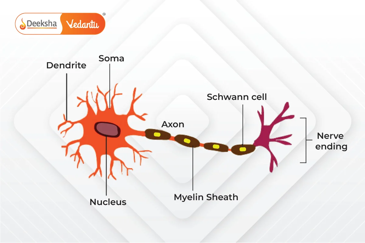
- Cell Body (Soma): Contains the nucleus and other organelles. It is responsible for maintaining the neuron’s health and function.
- Dendrites: Short, branch-like extensions that receive electrical signals from other neurons or sensory receptors.
- Axon: A long, thin fiber that carries the electrical signal away from the cell body towards other neurons or muscles.
At the end of the axon, there are synaptic terminals that release neurotransmitters, which allow communication between neurons or between a neuron and an effector (muscle or gland).
Key Features of Neurons:
- Myelin Sheath: In many neurons, the axon is covered by a myelin sheath, which acts as an insulating layer. It speeds up the transmission of electrical impulses along the axon. The myelin sheath is produced by Schwann cells and is interrupted at intervals by nodes called Nodes of Ranvier, where the nerve impulses “jump,” speeding up conduction.
- Synapse: A synapse is the gap between the axon terminal of one neuron and the dendrite of another neuron or an effector cell. The electrical impulse is converted into a chemical signal at the synapse. Neurotransmitters are released into the synapse and bind to receptors on the adjacent cell, transmitting the signal.
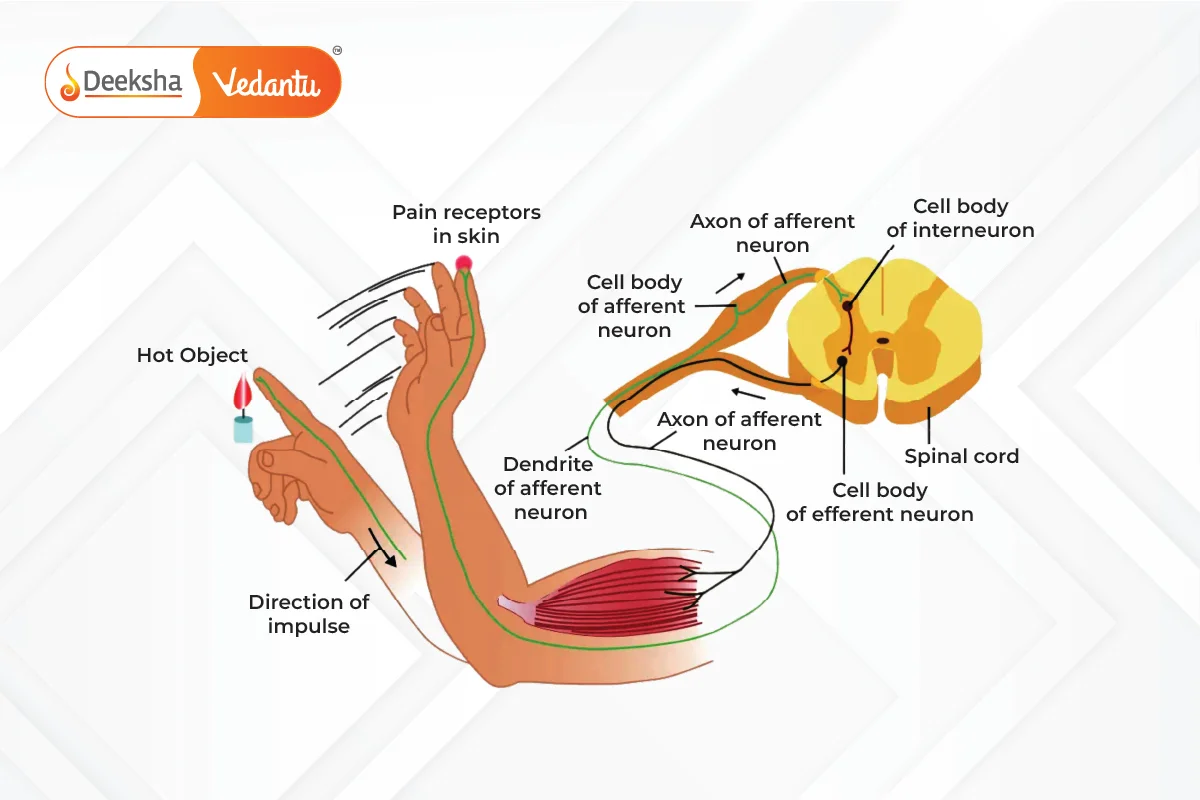
Example: When you touch a hot object, sensory neurons in your skin detect the heat and send a signal to the CNS. The brain processes this information and sends a response through motor neurons to move your hand away from the hot object.
Transmission of Nerve Impulses
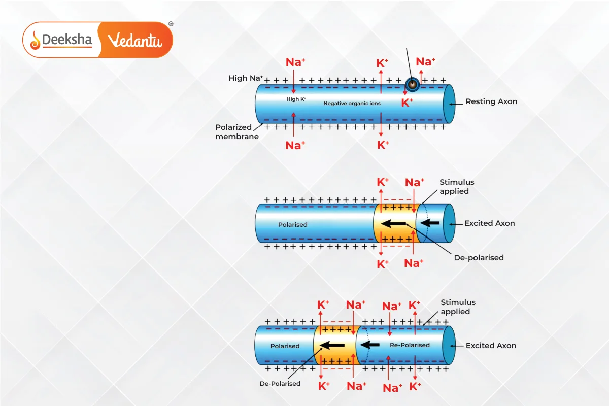
The transmission of nerve impulses involves the movement of electrical signals across neurons. This process is called action potential and involves the following stages:
- Resting Potential: When a neuron is not transmitting an impulse, it is in a state of resting potential. In this state, the inside of the neuron is negatively charged compared to the outside, due to the unequal distribution of ions (mainly sodium and potassium).
- Depolarization: When a neuron receives a stimulus strong enough to trigger an impulse, sodium channels open, allowing sodium ions to rush into the neuron. This causes the inside of the neuron to become positively charged, generating an action potential.
- Repolarization: After the action potential passes, potassium channels open, allowing potassium ions to exit the neuron, restoring the negative charge inside the neuron.
- Refractory Period: After an impulse has passed, there is a short period during which the neuron cannot generate another action potential. This ensures that the nerve impulse travels in one direction only.
Once the electrical signal reaches the end of the axon, it triggers the release of neurotransmitters into the synaptic cleft. These chemicals diffuse across the synapse and bind to receptors on the next neuron, generating a new electrical impulse.
Reflex Actions and the Reflex Arc
Reflex actions are quick, automatic responses to stimuli that do not require conscious thought. They help protect the body from harm by allowing it to respond rapidly to dangerous stimuli, such as pulling your hand away from a sharp object.
Reflex Arc
A reflex arc is the pathway through which a reflex action occurs. It involves the following components:
- Receptor: Detects the stimulus (e.g., pain, temperature, pressure) and initiates the impulse.
- Sensory Neuron: Transmits the impulse from the receptor to the spinal cord.
- Interneuron (Relay Neuron): Located in the spinal cord, the interneuron processes the impulse and transmits it to the motor neuron.
- Motor Neuron: Transmits the impulse from the spinal cord to the effector (muscle or gland).
- Effector: The muscle or gland that responds to the stimulus (e.g., contracting a muscle to pull your hand away).
Example: When you accidentally step on a sharp object, the pain receptors in your foot send an impulse through sensory neurons to the spinal cord. The spinal cord processes this information and sends a response through motor neurons, causing the muscles in your leg to contract and withdraw your foot from the sharp object.
Importance of Reflex Actions:
- Speed: Reflex actions are faster than voluntary actions because they bypass the brain and are processed directly by the spinal cord.
- Protection: Reflexes help protect the body from harm by triggering quick responses to dangerous stimuli.
- Involuntary Nature: Reflex actions are involuntary and do not require conscious thought.
The Brain – The Control Center of the Nervous System
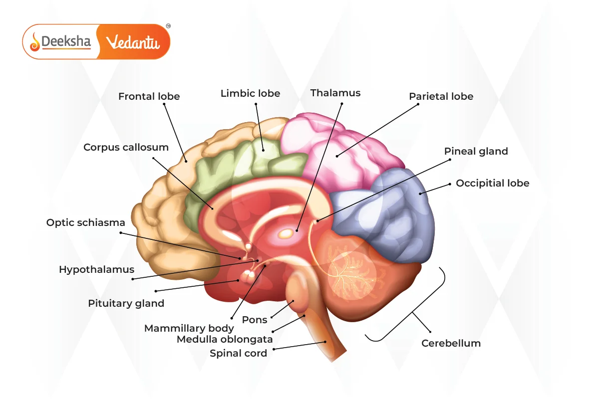
The brain is the main coordinating organ in the nervous system. It processes sensory information, stores memories, regulates emotions, and coordinates voluntary and involuntary actions. The brain is divided into three main parts:
Forebrain
- Cerebrum: The cerebrum is the largest part of the brain and is responsible for higher cognitive functions like thinking, reasoning, memory, and voluntary movements. It is divided into two hemispheres—left and right. Each hemisphere controls the opposite side of the body (the right hemisphere controls the left side of the body, and vice versa).
- Thalamus: Acts as a relay station, transmitting sensory information to the cerebrum.
- Hypothalamus: Regulates hunger, thirst, body temperature, and emotions. It also controls the endocrine system by regulating the secretion of hormones from the pituitary gland.
Midbrain
- The midbrain coordinates reflex responses related to vision and hearing. It also plays a role in regulating motor control and arousal.
Hindbrain
- Cerebellum: The cerebellum is responsible for coordinating balance, posture, and fine motor movements. It ensures that movements are smooth and precise.
- Medulla Oblongata: Controls vital involuntary functions such as heartbeat, respiration, and blood pressure.
- Pons: Connects the cerebellum to the rest of the brain and helps regulate breathing.
Protection of the Brain
The brain is protected by:
- Skull (Cranium): A hard bony structure that shields the brain from external injuries.
- Meninges: Three layers of membranes (dura mater, arachnoid mater, and pia mater) that surround and protect the brain and spinal cord.
- Cerebrospinal Fluid (CSF): A fluid that fills the spaces between the meninges and the brain cavities. It acts as a shock absorber and helps remove waste products from the brain.
Voluntary and Involuntary Actions
- Voluntary Actions: These actions are consciously controlled by the brain. They involve decision-making and planning in the cerebrum. Examples include walking, writing, or speaking.
- Involuntary Actions: These actions occur automatically without conscious control. They are controlled by the autonomic nervous system and the medulla oblongata. Examples include heartbeat, digestion, and breathing.
Key Difference:
- Voluntary actions require conscious thought and are under the control of the cerebrum, whereas involuntary actions are automatic and controlled by the brainstem and autonomic nervous system.
Practice Questions
Q1: Describe the structure and function of a neuron.
- Answer: A neuron consists of a cell body (soma), dendrites, and an axon. Dendrites receive electrical signals, the cell body processes them, and the axon transmits the signals to the next neuron or muscle through synapses.
Q2: Explain the process of nerve impulse transmission.
- Answer: A nerve impulse is generated when a neuron is stimulated. The impulse travels as an electrical signal along the axon, triggering the release of neurotransmitters at the synapse. These chemicals cross the synaptic gap and bind to receptors on the next neuron, transmitting the signal.
Q3: How does a reflex arc function?
Answer: A reflex arc involves receptors detecting a stimulus, sensory neurons transmitting the signal to the spinal cord, and motor neurons delivering the response to an effector (e.g., muscle contraction to pull away from danger).
FAQs
Reflex actions are faster because they are processed by the spinal cord and do not involve the brain. This allows the body to respond quickly to harmful stimuli without conscious thought.
The CNS (Central Nervous System) consists of the brain and spinal cord, which process and coordinate information. The PNS (Peripheral Nervous System) consists of nerves that connect the CNS to the rest of the body, carrying sensory and motor signals.
The nervous system detects changes in the environment (stimuli) through sensory receptors, processes the information in the brain and spinal cord, and generates an appropriate response through motor neurons.




Get Social