
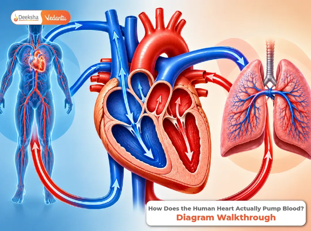


The heart is a fist-sized muscle that beats about 100,000 times a day without being asked, and it is one of the most reliable diagram questions in Class 10 Biology. Students lose marks on it for the same few reasons: they swap the sides, they forget the valves, or they cannot say in which order the chambers fill. All of that becomes simple once you follow the blood and not the labels.
This guide walks through one heartbeat, step by step, then through the double circulation, and finally through the drawing. For the other diagrams of this chapter, see the guide to the most commonly asked diagrams in life processes, and for the wider picture of how the heart fits with the other organs, the life processes comparison table.
Meet the Pump
The heart is made of a special muscle that contracts and relaxes rhythmically on its own. It sits in the chest, slightly to the left, between the lungs, and it does one job: it pumps blood through the blood vessels so that every cell receives oxygen and food and gives up carbon dioxide and wastes.
Why does it need four chambers? Because the blood has two kinds, and the heart needs to keep them apart. Oxygen-rich blood comes from the lungs and must go to the body. Oxygen-poor blood comes from the body and must go to the lungs. A single chamber would mix them, but a four-chambered heart keeps the two streams separate, which is exactly what a warm-blooded animal needs.
For a closer look at the organ itself, read these notes on the human heart and this piece on the structure and function of the heart.
Four Chambers, Four Valves
The upper two chambers are the atria (singular: atrium), which receive blood. The lower two are the ventricles, which pump it out. The ventricles have thicker walls than the atria, because they push the blood much farther.
| Chamber | Receives blood from | Sends blood to | Kind of blood |
| Right atrium | The body, through the vena cava | Right ventricle | Oxygen-poor |
| Right ventricle | Right atrium | Lungs, through the pulmonary artery | Oxygen-poor |
| Left atrium | The lungs, through the pulmonary veins | Left ventricle | Oxygen-rich |
| Left ventricle | Left atrium | The whole body, through the aorta | Oxygen-rich |
Blood must flow in only one direction, and valves make sure of that. Each valve is a set of flaps that opens one way only, like a door that swings in a single direction.
| Valve | Where it sits | What it prevents |
| Tricuspid | Between the right atrium and right ventricle | Backflow into the right atrium |
| Bicuspid (mitral) | Between the left atrium and left ventricle | Backflow into the left atrium |
| Pulmonary (semilunar) | At the exit to the pulmonary artery | Backflow into the right ventricle |
| Aortic (semilunar) | At the exit to the aorta | Backflow into the left ventricle |
One Heartbeat, Step by Step
A heartbeat is a cycle with a fixed order. It helps to think of it as a relay: first the atria push blood down, then the ventricles push it out.
- Filling. Blood flows into both atria, from the vena cava on the right and the pulmonary veins on the left.
- Atria contract. The atria squeeze, and the valves between the atria and ventricles open, so blood is pushed down into the ventricles.
- Ventricles contract. The ventricles squeeze strongly. The valves between the atria and ventricles close, which makes the first sound of the heartbeat, “lub”. The exit valves open, and blood is pumped out: the right ventricle sends it to the lungs and the left ventricle sends it to the body.
- Relaxation. The ventricles relax, and the exit valves snap shut, which makes the second sound, “dub”. The valves between the atria and ventricles open again, and the heart refills.
The doctor’s “lub-dub” is simply the sound of valves closing.
Double Circulation: Two Loops, One Heart
Blood passes through the heart twice in each complete round of the body, which is why this is called double circulation. The two loops are shown below.
Pulmonary circulation (heart ↔ lungs)
Right ventricle → Pulmonary artery → Lungs → Pulmonary veins → Left atrium
Systemic circulation (heart ↔ body)
Left ventricle → Aorta → Body → Vena cava → Right atrium
| Pulmonary circulation | Systemic circulation | |
| Goes to | The lungs | All other body organs |
| Pumped by | Right ventricle | Left ventricle |
| Blood in the artery | Oxygen-poor | Oxygen-rich |
| Blood in the vein | Oxygen-rich | Oxygen-poor |
| Purpose | Pick up oxygen and drop carbon dioxide | Deliver oxygen and food, and collect wastes |
The lungs are where the oxygen is picked up, as explained in why you breathe faster after running. Notice the one exception that students find odd: the pulmonary artery carries oxygen-poor blood, and the pulmonary vein carries oxygen-rich blood. The names depend on the direction of flow (arteries carry blood away from the heart, veins carry it to the heart), not on the oxygen content.
Why the Walls Are Different Thicknesses
The left ventricle has the thickest wall of all four chambers. The reason is the distance. The right ventricle only has to push blood to the nearby lungs, but the left ventricle has to push it through the whole body, from the brain to the toes, which needs much more pressure. A thicker muscle makes a stronger push. The atria have thin walls because they only push blood a short distance down into the ventricles.
Drawing the Heart: A Walkthrough
Use the same order every time, and the diagram becomes automatic.
- Draw the outline, a rounded shape that narrows at the bottom, and divide it into four chambers with a vertical and a horizontal line.
- Mark the sides correctly. The right side of the person’s heart goes on the left of your page, because you draw it as it faces you.
- Thicken the left ventricle wall.
- Add the vessels at the top: the vena cava entering the right atrium, the pulmonary artery leaving the right ventricle, the pulmonary veins entering the left atrium, and the aorta leaving the left ventricle.
- Draw the valves, small flaps between each atrium and ventricle, and at the exits.
- Label with ruled lines that do not cross, and add a title.
The usual slips are swapping the right and left sides, leaving the left ventricle wall the same thickness as the right, and forgetting that the vena cava and pulmonary veins both enter the atria, not the ventricles.
Heart Rate, Pulse and Pressure
A healthy adult’s heart beats about 70 to 80 times a minute at rest, and faster during exercise or fear. Each beat sends a wave along the arteries that you can feel as the pulse, for example on your wrist. Blood pressure is the force of blood on the walls of the arteries. It is usually written as two numbers, such as 120/80 mm of mercury, where the first is the pressure when the ventricles contract and the second is the pressure when they relax. Very high pressure over a long time can damage the heart and blood vessels, which is why it is checked in medical examinations.
Hearts Across the Animal World
| Animal group | Chambers | Circulation | Mixing of blood |
| Fish | 2 | Single: blood passes through the heart once per round | Not applicable |
| Amphibians and most reptiles | 3 | Double | Oxygen-rich and oxygen-poor blood mix partly |
| Birds and mammals | 4 | Double | Complete separation |
Birds and mammals keep their body temperature steady, which uses a lot of energy, so they need a heart that delivers oxygen as efficiently as possible. Complete separation of the two kinds of blood is what makes this possible.
The Heart in Your Textbooks
The heart is part of the transportation section of Life Processes, which is Chapter 5 in the CBSE Class 10 Science textbook for 2026-27 and Chapter 6 in the Karnataka SSLC textbook, according to published listings. Revise with these notes on transportation in humans and plants. In Class 11, the topic becomes body fluids and circulation, with the cardiac cycle in more detail, and for the NEET-level view, including blood pressure and ECG, see this guide to the heart mechanics for NEET. Confirm the current portions with your own board.
Frequently Asked Questions
Q1. How does the human heart pump blood?
The atria fill and contract to push blood into the ventricles. The ventricles then contract strongly, closing the valves between the atria and ventricles and opening the exit valves, so the right ventricle sends blood to the lungs and the left ventricle sends it to the body.
Q2. What is double circulation?
It is the passage of blood through the heart twice in one complete round of the body: once through the lungs (pulmonary circulation) and once through the rest of the body (systemic circulation).
Q3. Why is the wall of the left ventricle thicker?
It pumps blood to the whole body, which needs more pressure than pumping blood to the nearby lungs.
Q4. What is the role of valves in the heart?
Valves open in one direction only, so they keep blood flowing the right way and stop it from flowing backwards.
Q5. Which blood vessel carries oxygen-poor blood to the lungs?
The pulmonary artery. Arteries carry blood away from the heart, and in this case the blood is oxygen-poor because it has just come back from the body.
Follow the Blood
To master the heart, forget the labels for a moment and follow one drop of blood all the way round: vena cava, right atrium, right ventricle, lungs, left atrium, left ventricle, aorta, body, and back. Once you can say that path aloud, the diagram, the valves and the double circulation fall into place.









Get Social