
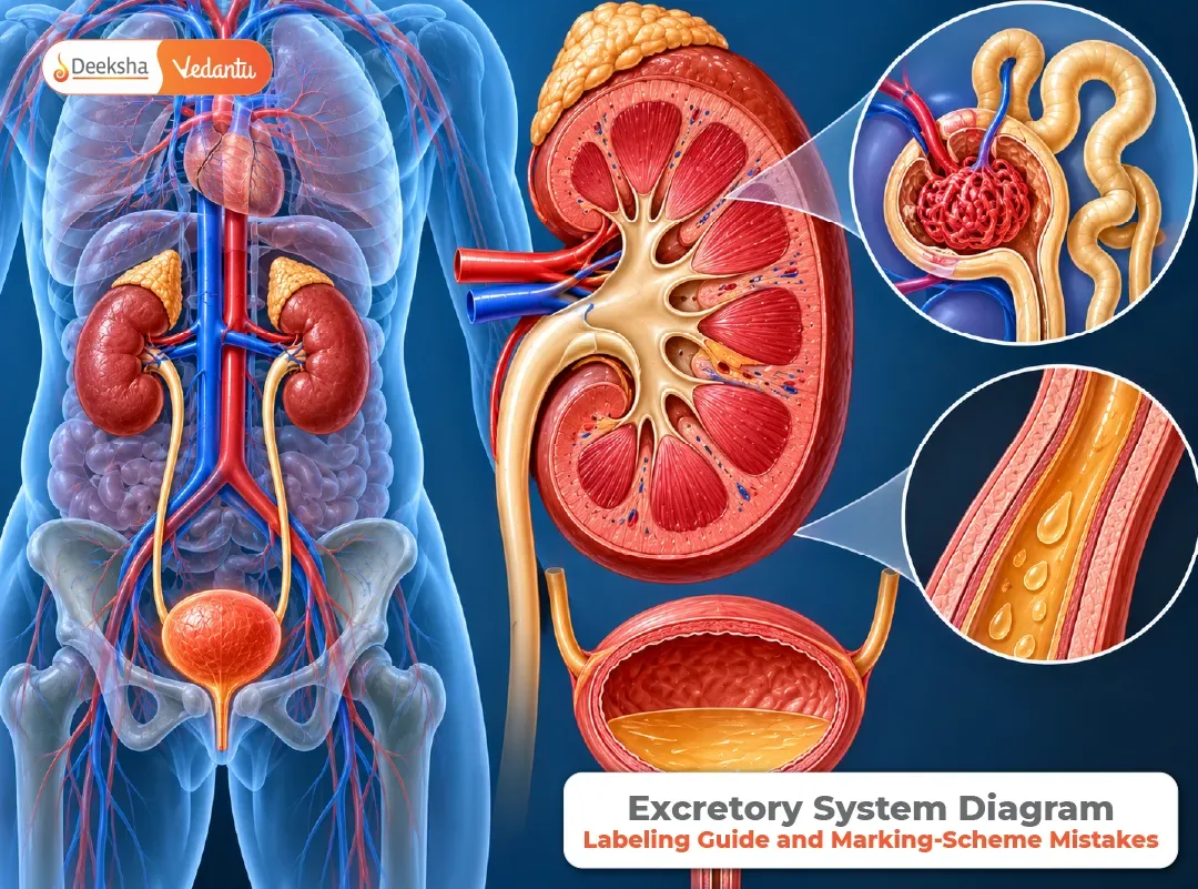


The excretory system is a favourite for diagram questions, because it is full of parts with similar-sounding names: ureter and urethra, renal artery and renal vein, Bowman’s capsule and glomerulus. Students who understand the process still lose marks by putting a label on the wrong tube.
This guide takes the two standard diagrams in turn, the whole system and a single nephron, and shows what to draw, what to label, and where the slips happen. It first explains the job of the system, because a diagram is far easier to draw when you know what each part is for. If you want the larger chapter context, see the life processes comparison table, the guide to the most commonly asked diagrams in life processes, and these notes on excretion.
What the System Does
As cells work, they produce wastes. The most important nitrogen-containing waste is urea, made from the breakdown of proteins. If urea and other wastes build up in the blood, they harm the body. The excretory system removes them, together with extra water and salts, and keeps the body’s water and salt balance steady.
The main organs are a pair of kidneys, which filter the blood and make urine, the ureters, which carry it away, the urinary bladder, which stores it, and the urethra, through which it leaves the body. The lungs and the skin also remove some wastes, carbon dioxide and sweat, but the kidneys do the main work.
Diagram One: The Whole System
What to draw
Start with the two kidneys, which are bean-shaped and lie on either side of the backbone in the abdomen. Then add the following in order.
- A large blood vessel coming into each kidney, the renal artery, and one leaving it, the renal vein.
- A thin tube from each kidney going down, the ureter.
- A sac where the two ureters meet, the urinary bladder.
- A single tube leaving the bladder, the urethra.
Labels to include
| Part | Where it is | Its job |
| Kidney | On each side of the backbone | Filters blood and forms urine |
| Renal artery | Enters the kidney | Brings blood with wastes to be filtered |
| Renal vein | Leaves the kidney | Takes filtered blood away |
| Ureter | From the kidney to the bladder | Carries urine down |
| Urinary bladder | Lower abdomen | Stores urine |
| Urethra | From the bladder to the outside | Passes urine out of the body |
The slips to avoid
The most common mistake is swapping ureter and urethra. A neat trick is that the ureter comes from the upper side, joining the kidney, while the urethra is the one at the end, taking urine out. There are two ureters and one urethra. Another slip is drawing the renal artery and vein with the same thickness. Draw the artery wider, since it carries blood under higher pressure.
Diagram Two: A Single Nephron
Each kidney has about a million tiny filtering units called nephrons. A nephron is where blood is actually filtered, so this is the diagram that shows how urine is made. You can read more about the details in this NEET-level guide to the nephron structure and filtration.
What to draw
- A cup-shaped structure at one end, the Bowman’s capsule.
- Inside the cup, a tight ball of blood capillaries, the glomerulus.
- A long, coiled tube leaving the cup, the renal tubule, with a blood capillary network wrapped around it.
- The tubule ending in a wider collecting duct, which carries urine towards the ureter.
- A small artery bringing blood to the glomerulus and a vein taking blood away from the capillaries around the tubule.
Labels to include
| Part | Job |
| Glomerulus | Capillary ball where blood is filtered |
| Bowman’s capsule | Cup that collects the filtrate |
| Renal tubule | Where useful substances are taken back |
| Capillaries around the tubule | Receive the substances taken back |
| Collecting duct | Carries urine to the ureter |
The slips to avoid
The glomerulus is inside the Bowman’s capsule, and not beside it. The tubule is not a straight pipe, it is long and coiled, and the capillaries around it are a part of the story, not decoration. A bare nephron with no blood vessels around it shows half the process.
How Urine Is Formed, Step by Step
The diagram only makes sense when you can read it as a process.
Blood → Glomerulus (filtration) → Bowman’s capsule → Renal tubule (selective reabsorption)
→ Collecting duct → Ureter → Urinary bladder → Urethra
- Filtration. Blood pressure forces water, glucose, amino acids, salts and urea out of the glomerulus into the Bowman’s capsule. Blood cells and large proteins stay behind.
- Selective reabsorption. As the filtrate passes along the tubule, useful substances such as glucose, amino acids, salts and much of the water are taken back into the blood in the surrounding capillaries.
- Collection. What remains, water with urea and excess salts, flows into the collecting duct. This is urine.
- Storage and release. Urine passes down the ureter into the bladder, where it is stored until it is released through the urethra. A ring of muscle at the exit controls the release.
The numbers show how selective this is. In a healthy adult, about 180 litres of filtrate are formed each day, but only a litre or two of urine leaves the body. Nearly all of the water and the useful substances are reabsorbed.
When the Kidneys Fail
If the kidneys stop working properly, wastes build up in the blood, and this can be life-threatening. One treatment is dialysis, using a machine that works as an artificial kidney. Blood from an artery is passed through a device in which wastes move out of the blood into a special fluid, and the cleaned blood is returned to a vein. Another treatment is a kidney transplant, in which a healthy kidney from a donor is placed in the patient. These show why understanding the nephron matters beyond the classroom: the machine copies what the nephron does.
A Labelling Checklist
- Are all six parts of the whole-system diagram present, with two ureters and one urethra?
- Is the glomerulus inside the Bowman’s capsule?
- Is the tubule coiled, with capillaries around it?
- Do the label lines end exactly on the part and avoid crossing?
- Is there a title under each diagram?
If you want to see how the same habits apply to another organ, the human heart diagram walkthrough uses the same method.
The Excretory System in Your Textbooks
Excretion is part of Life Processes, which is Chapter 5 in the CBSE Class 10 Science textbook for 2026-27 and Chapter 6 in the Karnataka SSLC textbook, according to published listings. For more, see the blog on the excretory system and life functions. In Class 11, the topic becomes excretory products and their elimination, which is part of NEET Biology. Confirm the current portions with your own board and the latest NMC document.
Frequently Asked Questions
Q1. What are the main parts of the human excretory system?
A pair of kidneys, two ureters, the urinary bladder and the urethra.
Q2. What is the difference between the ureter and the urethra?
The ureter is a tube that carries urine from each kidney to the bladder. The urethra is the single tube that carries urine from the bladder out of the body.
Q3. What is a nephron?
A nephron is the filtering unit of the kidney. It consists of a glomerulus, a Bowman’s capsule and a long renal tubule, and each kidney contains about a million of them.
Q4. How is urine formed?
Blood is filtered in the glomerulus into the Bowman’s capsule. As the filtrate passes along the tubule, useful substances and much of the water are taken back into the blood, and the rest becomes urine.
Q5. What is dialysis?
Dialysis is a treatment for kidney failure in which an artificial kidney removes wastes from the patient’s blood, and the cleaned blood is returned to the body.
Know the Job, Then the Label
Anatomical names are only difficult when they are separate from their jobs. The glomerulus filters, the tubule reabsorbs, the collecting duct collects, the ureter carries, the bladder stores and the urethra releases. Say that sentence aloud, and the diagram follows.











Get Social