Introduction
Did you know the human eye can distinguish nearly 10 million different colors? It’s one of the most remarkable organs in the body, allowing us to see the world in all its detail and vibrancy. It enables us to interpret shapes, motions, distances, and brightness. Understanding how the eye works and knowing the structure of the human eye is essential for students of Class 10 Science, especially for mastering topics in physics and biology.
This blog will walk you through the parts of the human eye, how vision works, and the scientific principles behind some common phenomena. We’ll also explore vision defects, real-life examples, and fun facts that make this topic both informative and exciting.
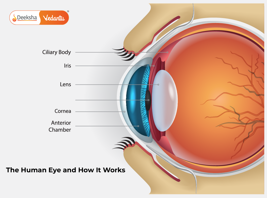
Structure of the Human Eye
The eye is shaped like a slightly flattened sphere and is about 2.5 cm in diameter. Despite its small size, it’s a complex organ with numerous intricate parts working in perfect harmony to create visual perception.
Learn more in detail here: Structure of Human Eye & Functioning
Major Parts of the Human Eye and Their Functions
1. Cornea
- Transparent, dome-shaped front surface of the eye
- Refracts (bends) light entering the eye and contributes to most of the eye’s focusing power
- Acts as the first lens in the eye’s optical system
2. Iris
- Colored portion surrounding the pupil
- Controls the diameter and size of the pupil, adjusting the amount of light entering the eye
- Works automatically in response to light intensity (pupil dilation and constriction)
3. Pupil
- Black circular opening in the center of the iris
- Appears black because light rays entering the pupil are absorbed by the tissues inside the eye
- Varies in size depending on light conditions and emotional responses
4. Lens
- Transparent, biconvex structure suspended behind the pupil
- Changes its shape to fine-tune the focus of incoming light (a process called accommodation)
- Works with the cornea to direct light precisely onto the retina
5. Retina
- Thin layer of tissue lining the inner surface at the back of the eye
- Contains two types of photoreceptor cells: rods (for night vision and peripheral vision) and cones (for color and sharp central vision)
- Converts light signals into electrical impulses for the brain
6. Optic Nerve
- Bundle of more than a million nerve fibers
- Transfers visual information from the retina to the visual cortex of the brain
- Responsible for creating the actual visual image in our minds
7. Aqueous and Vitreous Humors
- Fluids that fill the anterior (aqueous humor) and posterior (vitreous humor) chambers
- Maintain intraocular pressure, nourish internal eye structures, and help retain eye shape
How the Human Eye Works
Vision begins the moment light rays reflect off an object and enter the eye. The eye’s components function in a perfectly coordinated sequence to create a clear, focused image.
Step-by-Step Functioning
- Light enters through the cornea, which performs most of the focusing.
- The light then passes through the aqueous humor, reaching the pupil.
- The iris automatically adjusts the pupil size to control light entry.
- Light moves to the lens, which adjusts its curvature depending on object distance.
- The lens and cornea together focus light onto the retina.
- Retinal photoreceptors convert the light into electrical signals.
- The optic nerve transmits these signals to the brain.
- The brain interprets these signals to produce the image we perceive.
Fun fact: The image formed on the retina is inverted. It’s the brain that flips it right side up!
Explore further: The Human Eye
Real-Life Examples of Eye Functioning
Our understanding of the human eye becomes clearer when we relate it to daily life and familiar objects.
- Camera Analogy: Just like a camera, the eye uses a lens system (cornea and lens) to focus light onto a photosensitive surface (retina). The film or sensor in a camera is like the retina.
- Bright Sunlight Reaction: The iris automatically narrows the pupil to prevent excess light from entering, protecting the retina.
- Walking into a Dark Room: Initially, you see almost nothing, but after a while, your pupils dilate and rod cells in the retina adapt to the low light.
- Mirror Reflex in Eye Exams: During retinoscopy, doctors examine how light reflects from the retina to detect issues with vision.
- Red-Eye in Photos: Caused by camera flash reflecting off the retina, showing the blood vessels behind it.
Color Perception: The Colorful World
The world as we know it is full of colors, and our eye’s ability to detect and interpret color is due to specialized cells in the retina.
Understanding Colors
- Colors are perceived based on the wavelengths of visible light.
- Three types of cone cells in the retina respond to red, green, and blue wavelengths.
- Our brain processes the signals from these cones to interpret thousands of color shades.
- When an object reflects multiple wavelengths, cones send a combination of signals, creating blended colors.
- Color Blindness: A condition where one or more types of cones are missing or defective. Common in males, it affects the ability to differentiate certain colors, especially red and green.
More on this topic: The Human Eye and the Colourful World
Dispersion of Light and Eye Perception
One of the key topics tied to eye function and optics is dispersion—the process by which white light splits into its component colors.
- Example: When white light passes through a glass prism, it separates into seven colors—violet, indigo, blue, green, yellow, orange, and red (VIBGYOR).
- This happens because each color bends by a different amount when passing through the prism.
- Rainbows are a natural demonstration of dispersion. Sunlight disperses through raindrops, forming a spectrum.
- The cones in our eyes detect each color separately, helping us perceive the full range of visible light.
Explore the physics here: Dispersion of White Light by a Glass Prism
Frequently Asked Questions
1. What is the structure of the human eye?
The eye includes the cornea, iris, pupil, lens, retina, optic nerve, and fluids that nourish and support internal structures. These work together to convert light into visual signals.
2. How does the eye work?
The cornea and lens focus light onto the retina. The retina converts the light into signals, which are transmitted to the brain by the optic nerve. The brain interprets these to form visual images.
3. What are the parts of the eye and their functions?
Each part has a distinct function: the cornea and lens focus light, the iris regulates light entry, the retina processes it, and the optic nerve sends data to the brain.
4. Why do we see colors?
The cones in the retina detect different wavelengths of light. The brain processes these to produce the wide range of colors we see.
5. What causes red-eye in photos?
It’s due to light from the camera flash reflecting off the retina’s blood-rich layer.
Defects of Vision and Their Correction
Despite the eye’s precision, many people experience vision problems due to structural or functional issues. Fortunately, most of these defects can be corrected.
Tip: Including visuals or diagrams helps explain how lenses correct light paths and improve focus.
1. Myopia (Nearsightedness)
- Distant objects appear blurry
- Cause: Elongated eyeball or overly curved lens
- Light focuses in front of the retina
- Correction: Use of concave lenses to diverge light rays before they enter the eye
2. Hypermetropia (Farsightedness)
- Nearby objects appear blurry
- Cause: Short eyeball or flat lens
- Light focuses behind the retina
- Correction: Use of convex lenses to converge light rays
3. Presbyopia
- Age-related vision decline (usually after age 40)
- Difficulty in focusing on nearby objects due to reduced lens elasticity
- Correction: Bifocal or progressive lenses that aid both near and distant vision
4. Astigmatism
- Blurred or distorted vision at all distances
- Cause: Irregular curvature of the cornea or lens
- Correction: Cylindrical lenses or toric contact lenses designed to counter the uneven curvature
5. Night Blindness
- Inability to see well in low light
- Often caused by vitamin A deficiency or rod cell dysfunction
- Prevention: Diet rich in vitamin A (carrots, leafy greens)
Read in-depth here: Defects of Vision and Their Correction
Conclusion
The human eye is an extraordinary biological camera. From regulating light intake to processing complex images and detecting color, it plays an indispensable role in how we interact with the world. Understanding how it works not only helps in scoring better in exams but also deepens appreciation for human biology and health.
Whether it’s observing a rainbow, reading a book, or navigating in the dark, every experience is shaped by this small yet powerful organ. With proper knowledge, we can take better care of our eyes and make informed decisions about vision correction and eye health.
For more lessons, diagrams, and visual explanations on this topic, visit Deeksha Vedantu’s curated physics resources.
Table of Contents


Get Social