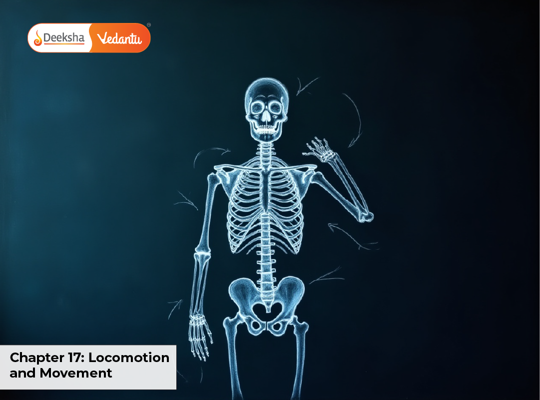
The chapter Locomotion and Movement typically contributes 2–3 direct questions in NEET (amounting to 8–12 marks), but its concepts can also be integrated into assertion–reason questions and diagram-based items. Students must master:
- All types of movement, with clear functional distinctions and examples
- Skeletal muscle ultrastructure and the complete sliding filament theory sequence
- Classification and examples of joints with movements permitted
- Locations and functions of major bones and muscles
- Common disorders such as arthritis, osteoporosis, muscular dystrophy, and their physiological basis
Many NEET questions are based on visual interpretation of sarcomere structure, sequence of muscle contraction events, and joint classification diagrams, making diagram practice essential.
Introduction to Locomotion and Movement
Movement refers to any change in the position or orientation of a body part. It is a hallmark of living organisms, occurring in forms ranging from microscopic intracellular transport to large-scale whole-body displacement. This includes activities like the streaming of cytoplasm, beating of cilia, and contraction of muscles.
Locomotion is a specialized category of movement that results in a change in the position of the organism as a whole from one place to another. Examples include walking, swimming, flying, running, and crawling. Locomotion often requires integrated action of the skeletal and muscular systems and is crucial for foraging, escaping predators, and seeking mates.
Key Differences:
- All locomotions are movements, but not all movements qualify as locomotions.
- Example: The rhythmic contraction of the heart is a movement but not locomotion; walking from one location to another involves both movement and locomotion.
- Locomotion generally involves higher energy expenditure and coordination of multiple organ systems compared to localized movements.
Types of Movement
- Amoeboid Movement
- Involves pseudopodia (extensions of cytoplasm)
- Example: Amoeba, leucocytes (WBCs)
- In humans: Macrophages and neutrophils use amoeboid movement for phagocytosis.
- Ciliary Movement
- Coordinated beating of cilia.
- Examples: Movement of ova in female reproductive tract, movement of mucus in respiratory tract.
- Muscular Movement
- Caused by contraction and relaxation of muscle fibers.
- Involves skeletal, smooth, and cardiac muscles.
- Responsible for voluntary locomotion and many involuntary activities.
Muscles in Humans
There are three types of muscles:
| Type | Location | Control | Appearance | Function |
| Skeletal | Attached to bones | Voluntary | Striated | Movement of limbs, posture |
| Smooth | Walls of hollow organs | Involuntary | Non-striated | Movement of food, blood vessels |
| Cardiac | Heart walls | Involuntary | Striated, branched | Pumping blood |
Skeletal Muscle Structure
- Each muscle is made of muscle bundles (fascicles) surrounded by connective tissue.
- Muscle fiber (cell) is multinucleated and contains myofibrils.
- Myofibrils are made of actin (thin) and myosin (thick) filaments arranged in repeating units called sarcomeres.
Sarcomere Structure:
- Z line – Boundaries of a sarcomere.
- I band – Light band containing actin only.
- A band – Dark band containing both actin and myosin overlap.
- H zone – Myosin only region in the middle of A band.
- M line – Middle of sarcomere.
Sliding Filament Theory of Muscle ContractionProposed by Huxley and Niedergerke and Huxley and Hanson (1954).Steps:
- Nerve impulse reaches neuromuscular junction → releases acetylcholine.
- Depolarization spreads → Ca²⁺ released from sarcoplasmic reticulum.
- Ca²⁺ binds to troponin → moves tropomyosin → exposes myosin-binding sites on actin.
- Myosin heads bind to actin → power stroke → actin slides over myosin.
- ATP is hydrolyzed to detach myosin heads → cycle repeats.
NEET Tip: During contraction, A band remains same length, I band and H zone shorten.Skeletal System OverviewThe human skeleton has 206 bones divided into:
- Axial Skeleton (80 bones) – Skull, vertebral column, sternum, ribs.
- Appendicular Skeleton (126 bones) – Limbs, pectoral, and pelvic girdles.
Functions:
- Support and shape
- Protection of organs
- Muscle attachment
- Mineral storage (Ca, P)
- Blood cell production (in bone marrow)
Joints in HumansJoints are points where two bones meet. They are classified as:
- Fibrous (Immovable) – Sutures in skull.
- Cartilaginous (Partially movable) – Vertebral column.
- Synovial (Freely movable) – Shoulder, hip, knee, elbow.
Types of Synovial Joints:
- Ball-and-socket (hip, shoulder) – movement in all directions.
- Hinge (elbow, knee) – movement in one plane.
- Pivot (atlas-axis) – rotational movement.
- Gliding (carpals) – sliding movement.
- Saddle (thumb) – back-and-forth and side-to-side movement.
Locomotion in Humans
- Walking and running: Alternating leg movement coordinated by skeletal muscles and joints.
- Swimming: Combination of limb, trunk, and sometimes tail movement.
- Specialized movements: Climbing, throwing, dancing — all depend on muscular-skeletal coordination.
Disorders of Muscular and Skeletal System
- Myasthenia gravis: Autoimmune, causes muscle weakness.
- Muscular dystrophy: Progressive degeneration of muscles.
- Tetany: Low Ca²⁺, causes muscle spasms.
- Arthritis: Inflammation of joints (osteoarthritis, rheumatoid arthritis).
- Osteoporosis: Bone mass reduction due to hormonal imbalance or aging.
- Gout: Uric acid crystal deposition in joints.
Quick Revision Table
| Concept | Key Points |
| Types of movement | Amoeboid, Ciliary, Muscular |
| Muscle proteins | Actin, Myosin, Troponin, Tropomyosin |
| Sarcomere changes | A band constant, I band shortens |
| Skeleton parts | Axial (80), Appendicular (126) |
| Synovial joints | Ball-and-socket, Hinge, Pivot, etc. |
| Disorders | Myasthenia gravis, Arthritis, Osteoporosis |
Practice MCQs
- Which part of sarcomere shortens during contraction?
a) A band
b) I band
c) Z line
d) M line
Answer: b) I band - The neurotransmitter at neuromuscular junction is:
a) Dopamine
b) Acetylcholine
c) Serotonin
d) Adrenaline
Answer: b) Acetylcholine - Which joint is present between atlas and axis?
a) Hinge
b) Pivot
c) Ball-and-socket
d) Saddle
Answer: b) Pivot - The human skeleton has how many bones?
a) 206
b) 196
c) 216
d) 256
Answer: a) 206 - Which disease involves degeneration of articular cartilage?
a) Osteoarthritis
b) Myasthenia gravis
c) Gout
d) Rickets
Answer: a) Osteoarthritis - Which ion is essential for muscle contraction?
a) Na⁺
b) K⁺
c) Ca²⁺
d) Mg²⁺
Answer: c) Ca²⁺ - Which joint is the most freely movable?
a) Hinge
b) Ball-and-socket
c) Pivot
d) Gliding
Answer: b) Ball-and-socket - Which muscle type is multinucleated?
a) Cardiac
b) Skeletal
c) Smooth
d) None
Answer: b) Skeletal - Troponin and tropomyosin are associated with:
a) Myosin
b) Actin
c) Sarcoplasmic reticulum
d) Mitochondria
Answer: b) Actin - Which bone is part of the axial skeleton?
a) Femur
b) Sternum
c) Humerus
d) Radius
Answer: b) Sternum
FAQsWhat is the difference between movement and locomotion?Movement is any change in position of body parts, while locomotion results in a change of location of the organism.Why is Ca²⁺ important for muscle contraction?It binds to troponin, causing a shift in tropomyosin, exposing myosin-binding sites on actin filaments.Which is the longest bone in the human body?The femur (thigh bone).Why do elderly people suffer from osteoporosis?Due to decreased estrogen/testosterone levels and reduced calcium absorption.What is the function of synovial fluid?It lubricates joints, reducing friction and allowing smooth movement.ConclusionLocomotion and Movement brings together muscle physiology, skeletal anatomy, and neural control to explain how the human body achieves precise, energy‑efficient motion. For NEET, focus on three pillars: (1) muscle: sarcomere labeling, band changes during contraction, cross‑bridge cycle with the roles of Ca²⁺ and ATP; (2) skeleton and joints: axial vs appendicular bones and the movements permitted by each synovial joint with classic examples; (3) clinical angle: keyword features of common disorders (myasthenia gravis, osteoarthritis, osteoporosis, gout) and what they imply for movement.Last‑minute checklist for revision:
- Redraw the sarcomere and mark A, I, H, Z, and M precisely; note what shortens vs stays constant.
- Write the sliding filament sequence in 5 numbered steps (ACh → Ca²⁺ release → troponin–tropomyosin shift → power stroke → ATP‑dependent detachment).
- Memorize one hallmark movement for each synovial joint (e.g., shoulder—abduction/adduction; knee—flexion/extension; atlas–axis—rotation).
- Match muscle type to site and control (skeletal—voluntary; smooth—visceral; cardiac—myogenic and fatigue‑resistant).
- Do 10 mixed MCQs daily from this chapter, including at least two assertion‑reason items and one diagram question.
Mastering these high‑yield ideas, along with steady diagram practice and brief daily MCQ drills, will lock in the conceptual clarity and speed you need to convert questions from this chapter on exam day.







Get Social